The biological mechanisms of epilepsy
An epileptic seizure corresponds to abnormally prolonged electrical activity in a group of neurons in the cerebral cortex. The action potential or nerve impulse is an electrical message created by an inversion of positive and negative charges on either side of the neuron’s membrane, resulting from an exchange of ions between the cell and its environment (Fig B). At rest, the positive charges are outside the neuron (Fig A).

Under normal conditions, the role of each neuron is to receive, process and transmit the electrical message to the other neurons via the synapse using neurotransmitters (green spheres in the diagram).
The neuron that has transmitted the electrical signal then enters a phase of repolarisation, i.e. a reversal of charges between the outside and inside of the neuron, during which it is inactive (FigC).
In the case of an epileptic seizure, the neurons become hyperexcitable, i.e. a single stimulation results not in an action potential but in a succession, a train of repetitive action potentials with no rest period (FigD).
In idiopathic epilepsies, this neuronal hyperexcitability can be explained by mutations in the ion channels located on the neuron’s membrane, which allow the exchange of ions and therefore depolarisation and repolarisation. In general, the membrane becomes too permeable, preventing a return to a resting potential.
Hyperexcitable neurons form the epileptic focus. A distinction is made between focal epileptic seizures, which originate in a very delimited region of the brain, and generalised seizures, which are the result of a train of action potentials spread throughout the brain.
During attacks, hyperexcitability is very often accompanied by hypersynchrony, with several groups of neurons generating trains of action potentials at the same time and at the same rhythm, amplifying the intensity of the symptoms.
At the Paris Brain Institute
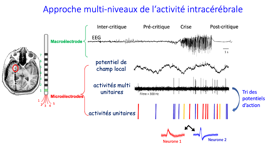
The “Cellular Excitability and Dynamics of Neuronal Networks” team, co-directed by Stéphane CHARPIER, Vincent NAVARRO & Mario CHAVEZ, is seeking to understand how the brain becomes epileptic (epileptogenesis), how it produces seizures and to identify the relationships between the abnormal electrical activity of a single neuron and the clinical signs observed in patients.

Implanting microelectrodes in the brain during intracerebral EEG scans of patients with treatment-resistant epilepsy is a new approach that makes it possible to monitor brain activity at the level of a few neurons during epileptic seizures, but also at a distance (inter-critical period) and shortly before seizures (pre-critical period). This information is acquired in the Epilepsy Unit, with the help of the Brain Institute’s CENIR-STIM platform, and the data is transferred directly to the ICM’s servers, where it is analysed by Professor Vincent Navarro’s team. This makes it possible to continuously monitor the activity of groups of neurons for days on end: the Institut du Cerveau is the only institute in France with such a recording procedure.
The brain consumes a large proportion of our daily energy intake. Synapses in particular, which link neurons together, consume a lot of energy. Every time neurons communicate with each other, a lot of energy is consumed. Unsurprisingly, not having enough energy to maintain neuronal communication has deleterious effects.
The aim of Jaime de Juan-Sanz’s team is to understand and identify the essential molecular mechanisms involved in maintaining the bioenergetics of synapses under normal conditions and to show a relationship between energy dysfunction and epileptic seizures.


