


How is post-stroke rehabilitation carried out?
“After a stroke and the appearance of a focal lesion, even if the underlying nerve tissue dies, there is a chance of recovery thanks to the brain’s plasticity. Using magnetic resonance neuroimaging (MRI) techniques, both static (observation of lesions) and dynamic (monitoring the activation of different brain regions during movement), we are studying the extent to which the different processes of cerebral plasticity are taking place in each patient: The surrounding tissues may take over the lost function, the secondary areas involved in movement programming may take part in movement control, or the contralateral areas (the healthy hemisphere, unaffected by the stroke) may take over, since we have 2 cerebral hemispheres. For example, when a person who has suffered a stroke shakes hands (when they can still do so), a different area of the brain is activated to that of a healthy person”.
Charlotte Rosso, neurologist specialising in stroke (neurovascular department, Pitié-Salpêtrière hospital), researcher in the “Mov’It: Movement, Investigations, Therapeutics” team. Normal and abnormal movement: physiopathology and experimental therapeutics” team led by Profs Marie VIDAILHET and Stéphane LEHERICY at the Institut du Cerveau.
Research projects at the Paris Brain Institute
- Depending on the location of the residual cerebral lesion following a stroke, the after-effects will be different. This is why it is important to know the structure of cerebral vascularisation. Nicolas RENIER’s Structural Network Dynamics team at the Institut du Cerveau aims to map this vascularisation in 3 dimensions in humans. A first step has been taken with the complete mapping of cerebral vascularisation in an experimental model.
- From assessing the after-effects of the acute phase to rehabilitation in “real-life” conditions, six specialist carers, doctors and researchers from the Institut du Cerveau in Paris explain the issues involved in complex post-stroke care and the future prospects for improving their patients’ recovery.
Stroke-related projects
- The Brain Institute’s iCRIN (Clinical Research Infrastructure) dedicated to stroke, coordinated by Prof. Charlotte Rosso, aims to develop long-term, effective collaboration between the clinical departments of the Pitié-Salpêtrière hospital and the Institute’s research teams.
The iCRIN is also a stakeholder in a French national register of all thrombectomies performed, run by the Foch Hospital in Paris, in order to assess the benefits of this technique on a very large scale in real-life conditions. The aim of this data collection is to assess the benefits of this invasive treatment in terms of patient recovery.
- The ATTACK-AVC project, led by Pr Charlotte ROSSO and Fabrizio DE VICO FALLANI of the “ARAMIS – Algorithms, models and methods for images and signals of the human brain” team, aims to characterise the profile of patients recovering from a stroke using parameters such as Magnetic Resonance Imaging (MRI) and Electroencephalography (EEG),
- The SPAST project, led by Jean-Charles LAMY and Prof. Charlotte ROSSO, is a therapeutic trial being conducted in collaboration with the start-up PATHMAKER, to treat hypertonia (involuntary muscle contractures) of the muscles of the lower limbs following a stroke, in order to improve patients’ walking by non-invasive electrical stimulation of the spinal cord.
- The GAIN project, the result of a partnership with the MINDMAZE start-up incubated at the Institut du Cerveau, focuses on the development and evaluation of the effect of augmented reality physiotherapy on post-stroke recovery. In France today, there are not enough physiotherapy resources to provide sufficient rehabilitation for stroke patients. Yet the dose of rehabilitation is essential for recovery: the greater the dose, the better the recovery. The aim is to find out whether physiotherapy, carried out autonomously by the patient via an augmented reality interface, produces good results in terms of recovery, and to identify which patients benefit most from it by means of brain mapping.augmented reality physiotherapy consists of making the patient imagine that he is performing a gesture, such as raising his arm, when he is physically incapable of doing so. The neural circuits are then brought into play and re-educated even though the gesture is not physically performed.
These three projects ended in 2021 and are no longer recruiting patients. Analyses are ongoing.
- The “PICNIC- Neuropsychology and functional neuroimaging” team, co-directed by Prof. Paolo BARTOLOMEO, is particularly interested in brain markers predicting recovery of visual neglect following a post-stroke lesion in the right cerebral hemisphere.
- Rehabilitation of post-stroke patients using therapeutic games – “Voracy Fish”.
Dr Marie-Laure WELTER, neurologist and researcher in the “Experimental Neurosurgery” team, coordinator of the LabCom Brain e-NOVATION at the Brain Institute, head of the deep brain stimulation activity in the Neurology Department and co-head of the “Walking/Balance/Posture/Movement/TMS and neuronavigation in humans” platform at the Paris Brain Institute.
The principle behind these therapeutic video games is that they can be played at home or in an institutional setting (at a practitioner’s home or in hospital). Patients can play alone, with a family member, or in a network with other patients. Playing the game remotely is monitored using a technological platform that enables various parameters of the patient’s motor behaviour to be collected and analysed by therapists, so that the video game can be adjusted to suit each patient and his or her progress. The idea behind the ‘therapeutic video game’ is both to overcome the difficulties encountered by patients in more conventional rehabilitation and to remedy their possible demotivation in the face of the repetitive aspect and the difficulty of accessing a therapist in the city. The advantage of this type of innovative treatment, which is designed to be fun, is that it can combine the different components of human behaviour: motor skills with body movements, cognition with the challenges and success objectives of the different levels of the game, while combining the motivational and emotional aspects with the fun aspect of the system. Brain e-NOVATION’s idea is to include these games – which are intended to complement rehabilitation by practitioners (therapists or physiotherapists) – in clinical trials in order to assess and demonstrate the effectiveness and value of this type of treatment and tool, whether for stroke with Voracy Fish or for other types of pathology. ”
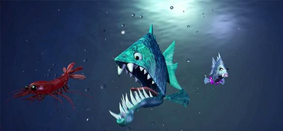
“Serious Game Voracy Fish from LabCom Brain e-NOVATION – partnership between the Institut du Cerveau – ICM and the GENIOUS group – Therapeutic game for functional rehabilitation of the upper limbs following a stroke © BRAIN e-NOVATION
- The BRO companion robot is the result of a collaboration between the Brain Institute’s CARELAB and the Physical and Rehabilitation Medicine (PRM) department run by Pr Pascale PRADAT at the Pitié Salpêtrière hospital.
Cerebral palsy patients have difficulty with activities of daily living, such as cooking. In this context, it is vital to improve the transfer of what has been learnt in hospital to the home (transfer of knowledge) using tools that are close to everyday life (ecological tools).
The aim of the BRO robot is to provide cognitive compensation for cerebral palsy patients during cooking activities. It encourages patients to regain their independence in hospital during therapeutic cooking sessions, and enables them to continue cooking at home. The specific BRO programme enables occupational therapists to define the difficulty of the task in order to adapt it to the patient’s cognitive profile. The programme consists of four criteria: memory, initiative, organisation and attention, which vary from level 1 to 4.
Depending on the criteria chosen, BRO offers one, two or three simultaneous written (and/or oral) instructions, reminders, images and/or videos, and aids adapted to the instructions.
- A NEW MATHEMATICAL MODEL OF BRAIN CONNECTIVITY AFTER STROKE
Researchers from the “ARAMIS – algorithms, models and methods for images and signals of the human brain” team, in collaboration with clinicians from the University of Padua (Italy), carried out a project on a cohort of stroke patients who underwent functional MRI 2 weeks, 3 months and 1 year after the stroke.
For each patient, the researchers modelled the increase in connection intensity in the damaged cerebral hemisphere, between the two hemispheres, and between the damaged region and its equivalent in the other hemisphere over time. The results of the neural network dynamics were correlated with clinical scores assessing each patient’s motor skills, vision, language, attention and memory.
The results of this study show that it is possible to reliably predict the recovery of language after a stroke, and constitute an innovative tool for identifying patient profiles likely to respond better to rehabilitation.
A new mathematical model of brain connectivity after stroke
- A PROMISING AVENUE FOR COMBINED REHABILITATION
Researchers and clinicians from the “MOV’IT: movement, investigations, therapeutics” team conducted a clinical trial on 27 stroke patients with a loss of fine motor skills in their hands (dexterity). The patients were treated with a series of concomitant transcranial magnetic stimulations of the cerebellum and motor cortex for 5 days and followed a physiotherapy programme targeting the upper limbs.
The results of this trial show that the combination of these different types of rehabilitation leads to an increase in activation (assessed by functional MRI) of the primary motor cortex (the brain region responsible for the planning, control and execution of voluntary muscle movements) in the injured hemisphere, as well as a significant improvement in dexterity that persists one month after the rehabilitation sessions.
Recordings of neuronal connections during and after the stimulation sessions have also enabled us to gain a better understanding of the mechanisms involved in this recovery, which will make it possible to adapt these therapies for optimum recovery.
- DEXTRAIN, A START-UP LOCATED AT THE BRAIN INSTITUTE FROM DECEMBER 2021
Dextrain is developing new digital tools for use in hospital or at home to assess and re-educate manual dexterity. The programmes can be adapted to each patient’s needs and level of performance, to provide continuous, adapted, fun and motivating re-education.
Based on knowledge of neuro-rehabilitation, a range of visuo-motor and audio-motor training exercises are designed to optimise recovery of the key components of dexterity and to encourage recovery, thereby contributing to independence in daily activities and improving quality of life.