


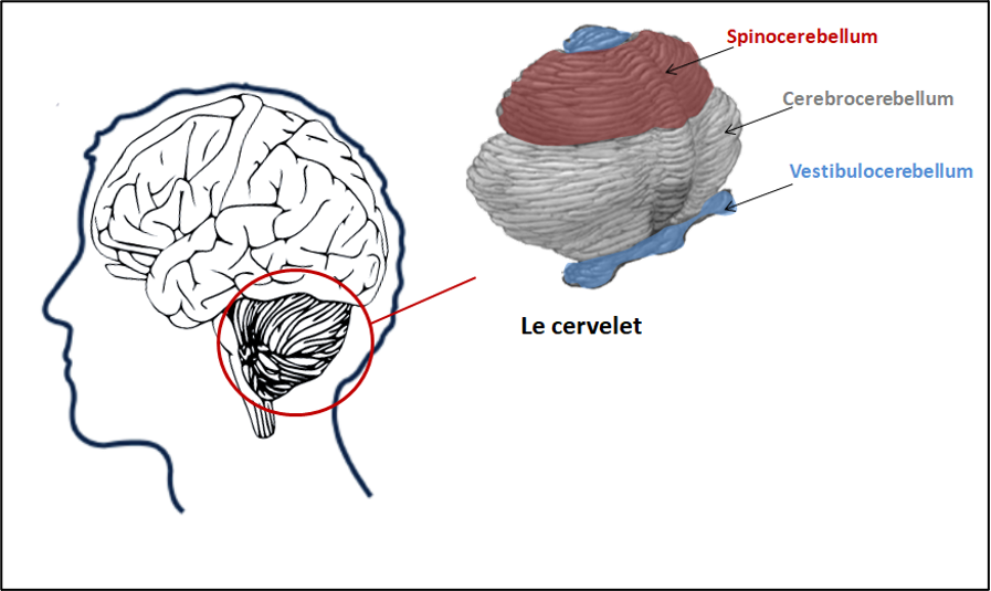
The cerebellum is made up of 3 parts:
- the vestibulocerebellum (or archicerebellum), which controls balance and eye movements
- The spinocerebellum (or vermis), which coordinates movements of the trunk and lower limbs, such as posture and walking.
- The cerebrocerebellum (or lateral hemispheres), which controls the precise and rapid movements of the upper limbs.
There are 3 main types of cerebellar ataxia:
- Ataxias due to congenital malformations of the central nervous system, particularly the cerebellum. Symptoms appear in childhood and do not progress very far.
- Acquired ataxias resulting from cerebral lesions, inflammation or prolonged and recurrent exposure to toxic substances.
- Hereditary ataxias due to genetic mutations that can be transmitted from generation to generation in a dominant or recessive mode.
Acquired ataxias
These pathologies have a sudden onset with no progression, and are mostly due to damage to the cerebellum following another disease or trauma.
A tumour, a cyst, a vascularisation disorder such as a stroke or haematoma, or a multiple sclerosis lesion, if located in or near the cerebellum, can cause symptoms of ataxia.
Similarly, an infection affecting the brain, such as HIV, prion disease or leukoencephalopathy, can sometimes lead to the appearance of ataxia symptoms.
Patients with acute vitamin B12 or E deficiencies, hyper- or hypo-thyroidism often present with cerebellar disorders.
Finally, prolonged and recurrent exposure to toxic substances such as alcohol, lead and organic solvents may be the cause of the onset of ataxia.
In the case of inflammatory, metabolic and toxic ataxias, there is a rapid progression of symptoms after a sudden onset, which only ceases once the cause has been eliminated.
Cerebellar ataxias of genetic origin
Autosomal dominant ataxias
These diseases have an incidence of 1-5/100,000 people. They include cerebellar and spinocerebellar ataxias.
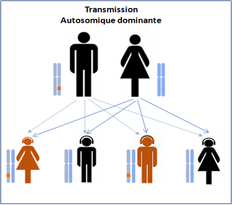
They appear on average around the age of 30 and are mainly due to genetic mutations, with at least 43 genes from the SCA (spino cerebellar ataxia) family located on different chromosomes.
In around 40% of patients, the gene responsible for the ataxia symptoms has not yet been identified.
Spinocerebellar ataxia type 3, with damage to the brain and spinal cord, due to a mutation in the SCA3 gene, is the most common (between 23 and 36% of familial cases).
At the Paris Brain Institute
The team co-led by Giovanni STEVANIN is particularly interested in spinocerebellar ataxias.
Institut du Cerveau – International Ataxia Awareness Day
Giovanni STEVANIN, INSERM/EPHE researcher and co-leader of the “Fundamental and Translational Neurogenetics” team
The team led by Dr Nathalie CARTIER, “Gene Therapy”, has discovered the crucial role of an enzyme in improving the symptoms of ataxia in an experimental model.
Autosomal recessive ataxias
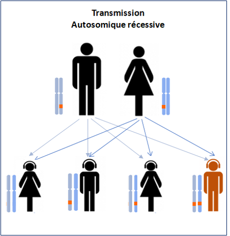
Autosomal recessive ataxias most often appear before the age of 25, although certain forms can appear before the age of 5.
A distinction is made between early-onset ataxias, Friedreich’s ataxia and ataxia telangiectasia.
-
Friedreich’s ataxia
This condition affects around 1,300 patients in France, with symptoms generally appearing between the ages of 7 and 14. This ataxia is due to mutations in the X25 gene, which codes for a protein called FRATAXIN, whose role is to regulate the quantity of iron in the mitochondria, a small structure essential to the survival of cells such as neurons.
When frataxin is mutated, it accumulates in the mitochondria, leading to dysfunction of the cerebellum. In this pathology, symptoms are essentially due to the death of neurons in areas of the spinal cord whose role is to inform the brain about body position, balance and motor skills.
At the Paris Brain Institute
The “Fundamental and Translational Neurogenetics” team, co-directed by Prof Alexandra DURR and Giovanni STEVANIN, is working on Friedreich’s ataxia.
In particular, this team has identified a neuropsychological profile of patients suffering from this ataxia.Ataxia telangiectasia
And is taking part in an international phase 2a therapeutic trial, FRAMES, designed to assess the efficacy and safety of MIN-102 in patients with Friedreich’s Ataxia.
https://institutducerveau-icm.org/fr/actualite/frames-therapeutique-lataxie-de-fredreich/
-
L’ataxie télangiectasie
The gene responsible for this disease is called ATM. It codes for a protein that “repairs” DNA, the molecule that carries genetic information. In particular, its role is to prevent cell death.
This ataxia generally begins around the age of 1 or 2. As with other ataxias, the symptoms observed are caused by an excess of neuronal death in the cerebellum, as the neurons are unable to ‘repair’ their DNA due to a lack of functional ATM protein.
The name telangiectasia comes from a particular symptom observed in patients and specific to this pathology: the dilation of small blood vessels in the eye, inside the ears, on the eyelids and in the folds of the elbows and knees.