


Several studies by Prof. Bruno Stankoff’s team “Remyelination in multiple sclerosis: from biology to clinical translation”, highlight new mechanisms of brain inflammation in multiple sclerosis, thanks to new imaging tools based on the combination of magnetic resonance imaging (MRI) and positron emission tomography (PET).
Using PET-MRI, Prof. Stankoff’s team has just published in the journal Radiology the results of a study of 97 MS patients and 44 healthy controls showing abnormalities (increased volume and inflammation) in the choroid plexuses of the patients. The choroid plexuses are structures located in the cerebral ventricles responsible for the production of cerebrospinal fluid, and act as a barrier between the nervous system and the immune system.
These choroidal plexus abnormalities were correlated with brain inflammation, indicating disease activity. These results open a new avenue for the application of imaging of this structure as a marker of the immune response in the brain and point to the choroid plexus as an important player in the pathophysiology of the disease.
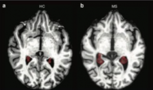 The choroïd plexuses (circled in red) of MS patients with a remitting form are larger (on the right) than healthy controls’ (on the left)
Work by the same team published in the journal Neurology, shows activation of innate immune cells associated with white matter micro-lesions in MS patients with worsening disability, and that this immune activation follows a gradient centered around the cerebral ventricles, which contain cerebrospinal fluid.
These results obtained thanks to the combination of PET targeting innate immune cells and MRI confirm the existence of a correlation between the activation of these cells around the ventricles, privileged areas of MS lesions in contact with cerebrospinal fluid (CSF), and the worsening of the disability in patients. This suggests that molecules contained in the CSF could worsen the inflammation present in the white matter of patients and thus promote a deleterious evolution of the disease, making these molecules candidates for future research into treatments.
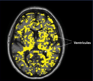 Activated innate immune cells in a patient with MS using PET-scan around ventricles.
Source
Structural and Clinical Correlates of a Periventricular Gradient of Neuroinflammation in Multiple Sclerosis. Poirion E, Tonietto M, Lejeune FX, Ricigliano VAG, Boudot de la Motte M, Benoit C, Bera G, Kuhnast B, Bottlaender M, Bodini B, Stankoff B. Neurology. 2021 Apr 6;96(14):e1865-e1875.
Choroid Plexus Enlargement in Inflammatory Multiple Sclerosis: 3.0-T MRI and Translocator Protein PET Evaluation. Ricigliano VAG, Morena E, Colombi A, Tonietto M, Hamzaoui M, Poirion E, Bottlaender M, Gervais P, Louapre C, Bodini B, Stankoff B. Radiology. 2021 Oct;301(1):166-177.
Positron emission tomogramphy in multiple sclerosis – straight to the target. Bodini B, Tonietto M, Airas L, Stankoff B. Nat Rev Neurol. 2021 Sep 20.