


The team of Paolo Bartolomeo at the Brain and Spine Institute (Institut du Cerveau – ICM), showed for the first time, that a brain lesion can “improve” visual perception in some patients, who are no longer subject to an optical illusion. These results illustrate the fact, essential but not well known, that normal vision is not a copy of the external world but instead an interpretation that the brain makes that functions to allow us to anticipate what is happening and to allow us to adapt. The study was published in the Journal of Neurophysiology.
Following a stroke that generates a lesion in the right hemisphere of the brain, some patients no longer pay attention to what happens in the left part of space; they suffer from “visual neglect” (or unilateral spatial neglect). They do not eat what is on the left half of their plate, they knock into walls on their left, they don’t shave or put on makeup on the left side of their face.
The team of Paolo Bartolomeo, Director of Research at INSERM and co-team leader at the Institut du Cerveau – ICM, in collaboration with the team of Patrick Cavanagh at the Psycology and Perception Laboratory (Paris Descartes University), showed that this brain lesion could “improve” the perception of these patients. Because of their attention deficits, these patients are not subjected to a visual illusion, usually very strong, when this takes place in their neglected space. In other words, they see in a more “real” fashion, more “precise” than people who do not have this brain lesion.
The test conducted consists of defining the placement of green and red lines that appear on a moving background. Look at them. Are they superimposed and horizontal? Is the green line on the top and the red line on the bottom? You probably see that the green line is on the top and the red line is on the bottom, while patients suffering from “visual neglect” see two superimposed horizontal lines.
We perceive that the lines are separate because of the moving background. Indeed, our brain integrates the movement and positions of the lines at the same time, as if it was trying to predict where the lines would be located is they followed the movement.
The patients, who are requested to pay attention to their left side, are not capable of integrating the movement from the background and the position of the lines. Therefore, they are not subject to the optical illusion and see the lines exactly as they are. Thus, our brain does not take a photo of reality but interprets it. What is the interest of this interpretation? It allows us to anticipate the outcome of a movement. When we play tennis, squash, or ping-pong, we need to predict the trajectory of the ball in order to adapt and to react to it. This is what our brain does when integrating the ball’s movement and in anticipating what the position of the ball will be when we hit it.
The team of Paolo Bartolomeo showed in this way that attention is fundamental for having this optical illusion, and when it does not work properly, the illusion is not longer present. Attention is therefore equally important for predicting the position of a moving target and having the time to react. The brain does not like to be surprised, and for that reason, it performs spatial and temporal integration that allows us to interpret what is going to happen. Brain lesions can damage this interpretation process in patients, leading to sight that is more “faithful” to external reality but less useful in daily life.
Our perceptions lead us to an interpretation of the world that is not the actual world but allows us to adapt. These results underline the important but poorly understand fact that normal vision is not a copy of the external world, but rather an interpretation of the world made by the brain. This interpretation allows us to generate adapted behavior and allows us to anticipate.
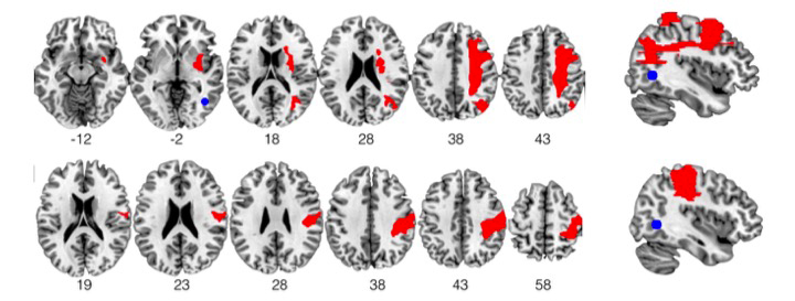
Lesions of the brain (in red) from two patients with visual neglect. The area for movement perception (in blue) do not have any lesion for these patients.