A paper published in the prestigious journal Brain by a team of the Institut du Cerveau – ICM institute link rapid eye movement sleep disorders and Parkinson’s disease. These results open the possibilities to early diagnose development of the Parkinson’s disease.
Parkinson disease is a progressive degenerative neurological disorder affecting 150 000 persons in France. It is characterized by the death of a neuronal population producing dopamine, a neurotransmitter, chemical messenger allowing transmission of informations between neurons. The diminution of the concentration of dopamine is the source of the development of the disease. Indeed, symptoms arise when 60-80% of these specific neurons disappear.
These last few years, researchers studied non-dopaminergic symptoms and more notably rapid eye movement sleep behavior disorder affecting 30 to 60% of patients. Rapid eye movement sleep is traditionally characterized by dreams and is associated with a strong neuronal activity, ocular movements and diminution of muscle tone. Some patients with Parkinson disease have rapid eye movement sleep behavior disorders with increased muscular tone which could lead to nocturnal violence. Rapid eye movement sleep behavior disorders could arise in any healthy subject whithout neurological disease. However, these persons present a higher risk to develop Parkinson disease or demencia. Daniel GarcÍa-Lorenzo in the team of Marie Vidailhet et Stéphane Lehéricy just highlighted the role of a part of the brain named the coeruleus/subcoeruleus complex in the blocking of muscle tone during eye rapid movement sleep.
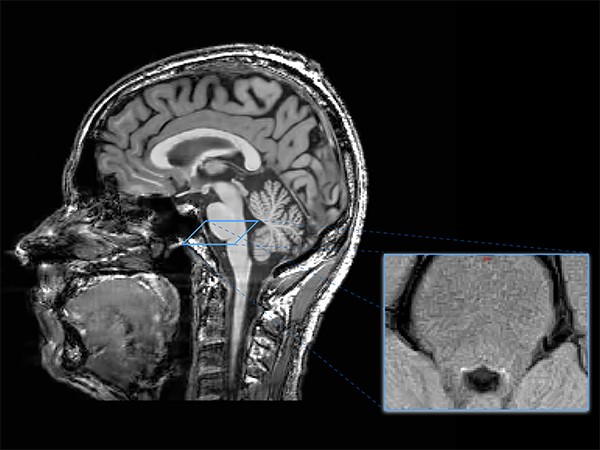
The team studied the integrity of this complex in patients with Parkinson’s disease using imaging approaches. They showed that patients with Parkinson’s disease have a reduced neuronal activity in the coeruleus/subcoeruleus complex associated with rapid eye movement sleep behavior disorder. Imaging datas combined with careful clinical evaluation as well as nocturnal video monitoring may be used as an early marker of non-dopaminergic Parkison’s disease pathology to predict the occurrence of Parkinson’s disease.

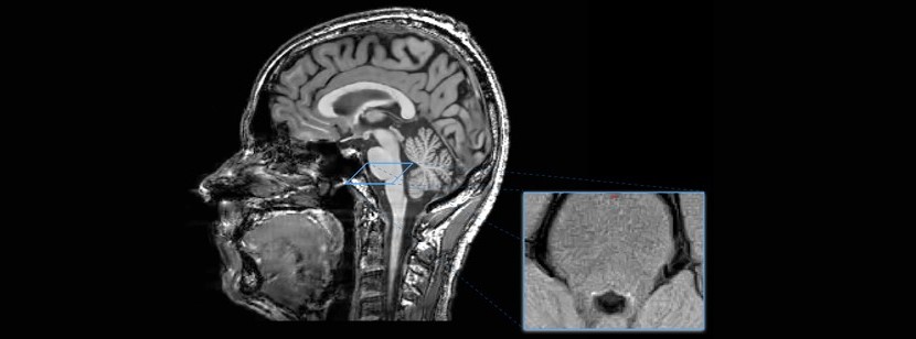
The coeruleus/subcoeruleus complex is visible in this new MRI sequence. This allowed us to study the relation between these small structures (in white on the image) and the muscle tone during rapid eye movement sleep.
References : Garcia-Lorenzo D, Longo-Dos Santos C, Ewenczyk C, Leu-Semenescu S, Gallea C, Quattrocchi G, Lobo PP, Poupon C, Benali H, Arnulf I, et al.: The coeruleus/subcoeruleus complex in rapid eye movement sleep behaviour disorders in Parkinson’s disease. Brain 2013, 136:2120-2129.


