


In an article recently published in the European Journal of Neurology, Vito Ricigliano (AP-HP), Benedetta Bodini (AP-HP/Sorbonne University) and their collaborators at the Paris Brain Institute, demonstrate the protective effect of myelin repair on the tissues surrounding lesions in patients with multiple sclerosis. This discovery underlines the potential of new therapeutic strategies and provides new elements to evaluate the efficacy of remyelinating drugs currently being tested.
How to prevent or reduce neuronal degeneration, which is the cause of clinical disability in multiple sclerosis (MS)? At present, doctors have treatments available to control the inflammatory component of the disease but are powerless to deal with the degenerative component.
“The study of experimental models has shown that myelin repair can protect the integrity of neurons and prevent neurodegeneration, which spreads from the demyelinating lesion all along the nerves that are not yet demyelinated but will degenerate directly,” explains Benedetta Bodini, neurologist and last author of the article.
In fact, in multiple sclerosis, the damage to the neurons is not only at the level of the myelin lesions visible on MRI, but extends to the regions surrounding them, the peri-lesional tissue. Researchers and clinicians at the Paris Brain Institute wanted to study whether spontaneous recovery of myelin – or remyelination – in the lesions could protect patients from microstructural damage to the surrounding tissue.

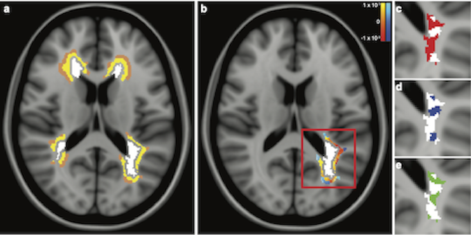
Lesions, perilesions and indices of myelin content change. a. Examples of lesional (white), proximal perilesional (yellow) and distal perilesional (brown) regions in the MRI of a patient with MS. b. White matter lesion (white) and its corresponding perilesions are represented with color maps, based on the delta of the mean diffusivity in each proximal and distal perilesional voxel from the first to the second time-point. c-e. Detail of the same white matter lesion of Fig. 1b, showing demyelinated voxels at baseline (red, c), dynamically demyelinating voxels over the follow-up (blue, d) and remyelinating voxels over the follow-up (green, e).
To do this, they monitored the amount of myelin in the lesions over time using PET-MRI and combined this with an assessment of the microstructural damage to the peri-lesional tissues using diffusion MRI, in 20 patients with multiple sclerosis. Their analyses were conducted at the single lesion level, i.e. over 500 lesions studied.
“We show for the first time in vivo in MS patients that remyelination protects not only the lesion but also the surrounding tissue. This result highlights the importance of coupling existing anti-inflammatory therapies with a remyelination strategy to protect all tissues, even those that appear normal” explains Vito Ricigliano, neurologist and first author of the study.
There is great heterogeneity among patients in their ability to remyelinate. In this study, the scientists also show that in the same patient, some lesions can repair themselves very well and others much less so, and that this difference is reflected in the damage to the peri-lesional tissue.
Clinical trials of remyelinating therapies are underway, notably in the team of Bruno Stankoff and Catherine Lubetzki at the Paris Brain Institute. Thanks to the use of PET-MRI, researchers will be able to study the effectiveness of the treatments, not only on the clinical signs, but also at the cellular level with the repair of myelin and the reduction of the microstructural damage to the surrounding tissues.
Source
Spontaneous remyelination in lesions protects the integrity of surrounding tissues over time in multiple sclerosis. Ricigliano VAG, Tonietto M, Hamzaoui M, Poirion É, Lazzarotto A, Bottlaender M, Gervais P, Maillart E, Stankoff B, Bodini B. Eur J Neurol. 2022 Feb 12.