It was a long-awaited event. On Sunday June 9, 2024, Paris Brain Institute welcomed an incredible piece of technology into its facilities: the latest generation MAGNETOM Terra X (Siemens Healthineers) 7T MRI. Teams have been doing a great deal of logistical and technical planning for the installation of this technology, which has been scheduled for a long time. We take you behind the scenes of a project that will be a milestone in the Institute’s history.
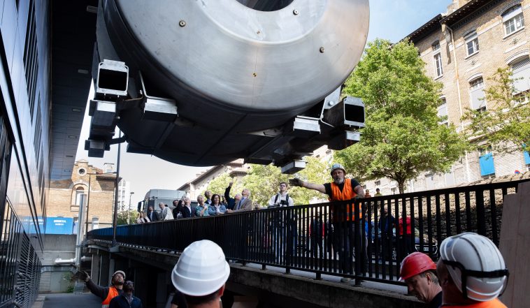
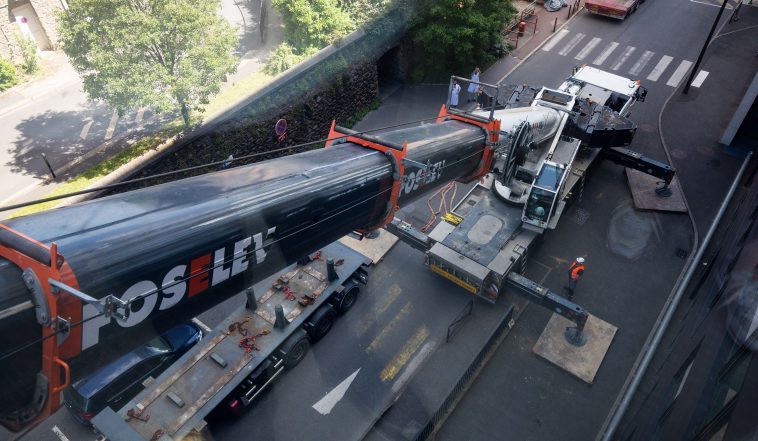
This is a magnet weighing at least twenty tons, made from a superconducting niobium-titanium alloy in a liquid helium bath at a very low temperature, to create a magnetic field of 7 Tesla, i.e. 140,000 times the Earth’s magnetic field. Installing this type of equipment in the heart of the 13th arrondissement is no mean feat.
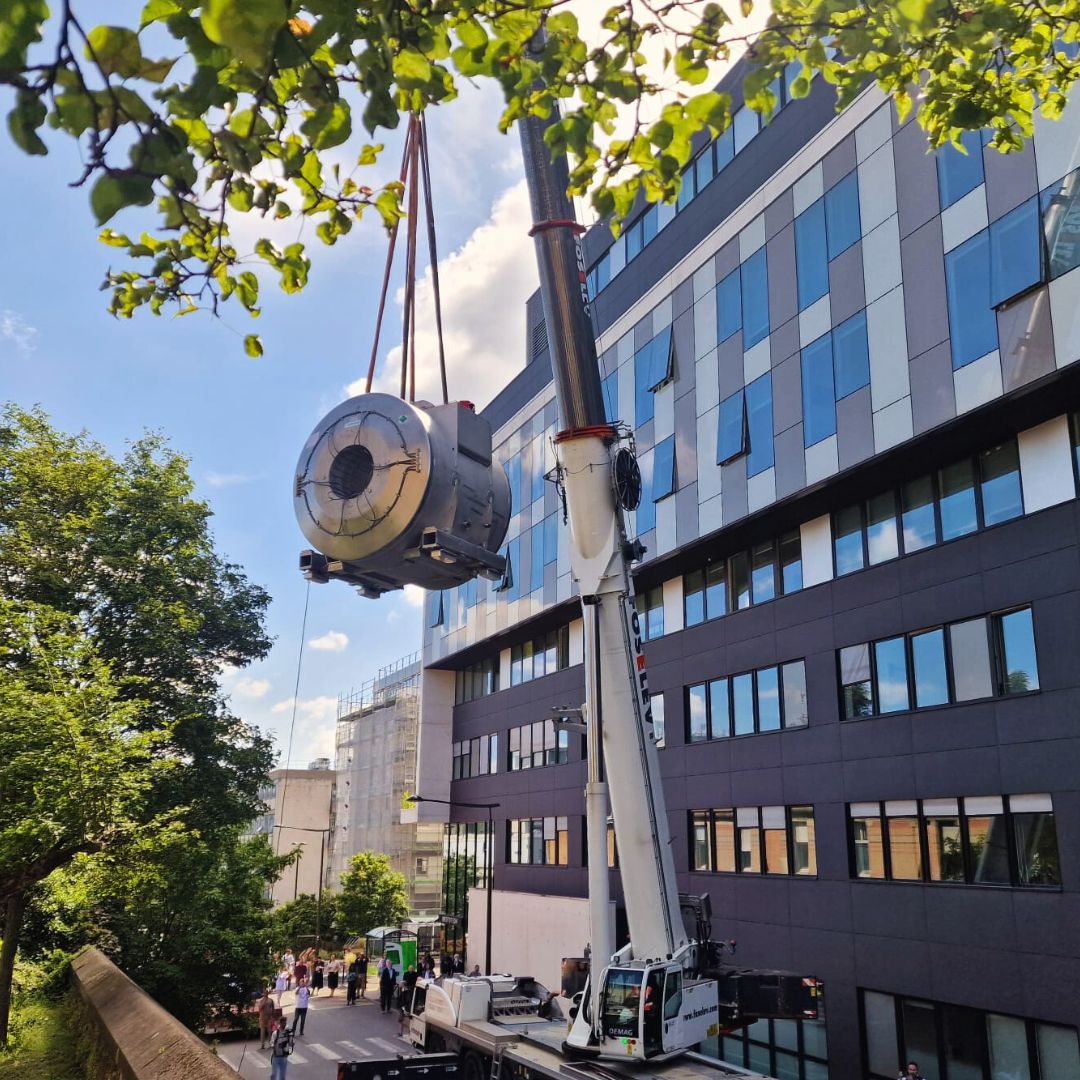
Arrival of the 7T MRI magnet outside the front of Paris Brain Institute
Operating the 7T MRI
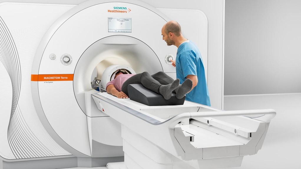
©Siemens
The magnet is key to the MRI’s operation. It creates a magnetic field with the strength of 7 Teslas – a unit of measurement established by Serbian physicist Nikola Tesla. By comparison, the Earth’s magnetic field is only 0.00005 Teslas. Just as the Earth’s magnetic field guides the needle of a compass North, the magnetic field of the MRI, known as the main field, allows the hydrogen atoms contained within the water molecules of the scanned person’s body to orient themselves along an axis parallel to the tube (shown by the red arrow on the diagram).
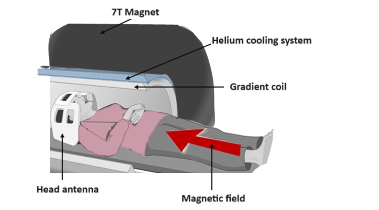
Magnetic pulses called radio frequencies, transverse to the main field, and generated by the gradient coils, modify the magnetic properties of the hydrogen atoms in a transient way. The return to equilibrium generates a magnetic signal registered by an antenna.
The way that the atoms “respond” to these pulses and the intensity of the MRI signal generated depend on the properties of the tissue in which the atoms are located. The MRI signal is recorded by a receiver reel and “translated” by the machine into 2D or 3D images.
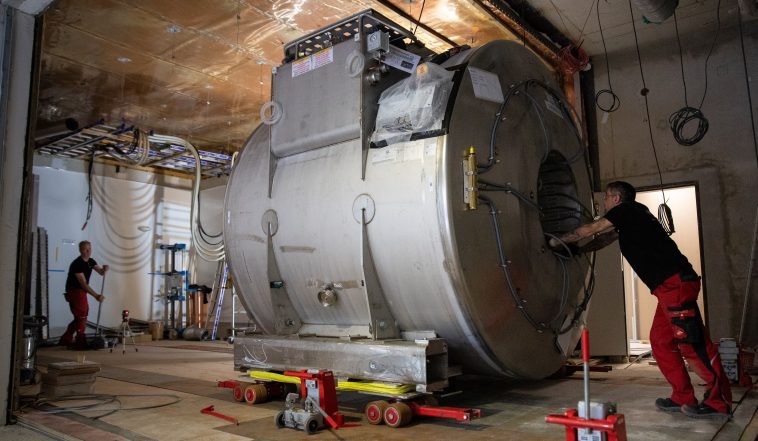
Building a fully hermetic room: a Faraday cage
To function correctly, there are certain constraints on an MRI machine’s installation (see above). The main magnetic field must be strictly limited to the room in which the MRI is located, for safety reasons, to avoid attracting or disrupting the operation of metal or electronic elements passing nearby.

Room housing the 7T MRI
To achieve this, the walls and floor of the room have been covered with shielding made from an alloy called mu-metal, made from 21% iron and 74% nickel. In addition, to produce a high-quality signal that can be used in medical and scientific imaging, the MRI must be protected from external electromagnetic interference by a Faraday cage made from copper foil.
The control and acquisition room, from which the radiographers control the machine, has a window overlooking the MRI room, fitted with hermetic glazing to shield it from the magnetic and electric fields.
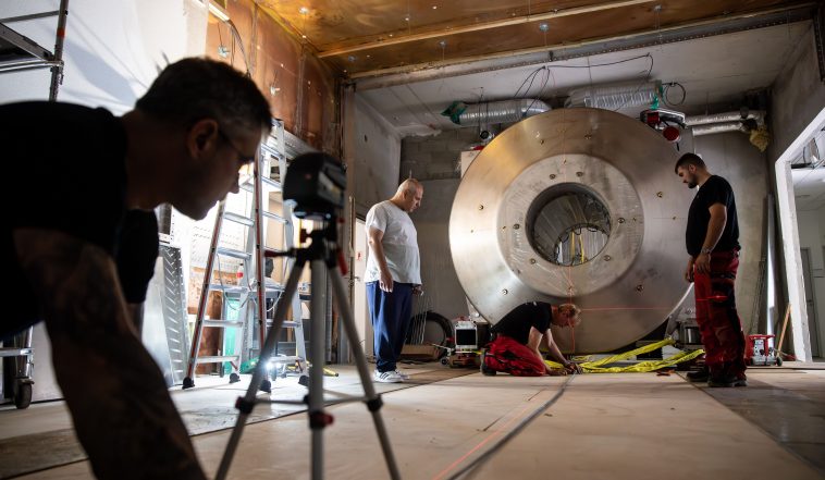
Entry into service and fine-tuning: one final hurdle before research use
The 7T MRI magnet installed at Paris Brain Institute is the latest generation of this technology: it is the lightest magnet of this strength in the world. It uses a zero helium evaporation system that recondenses helium vapors into liquid helium. The device has been given CE marking.
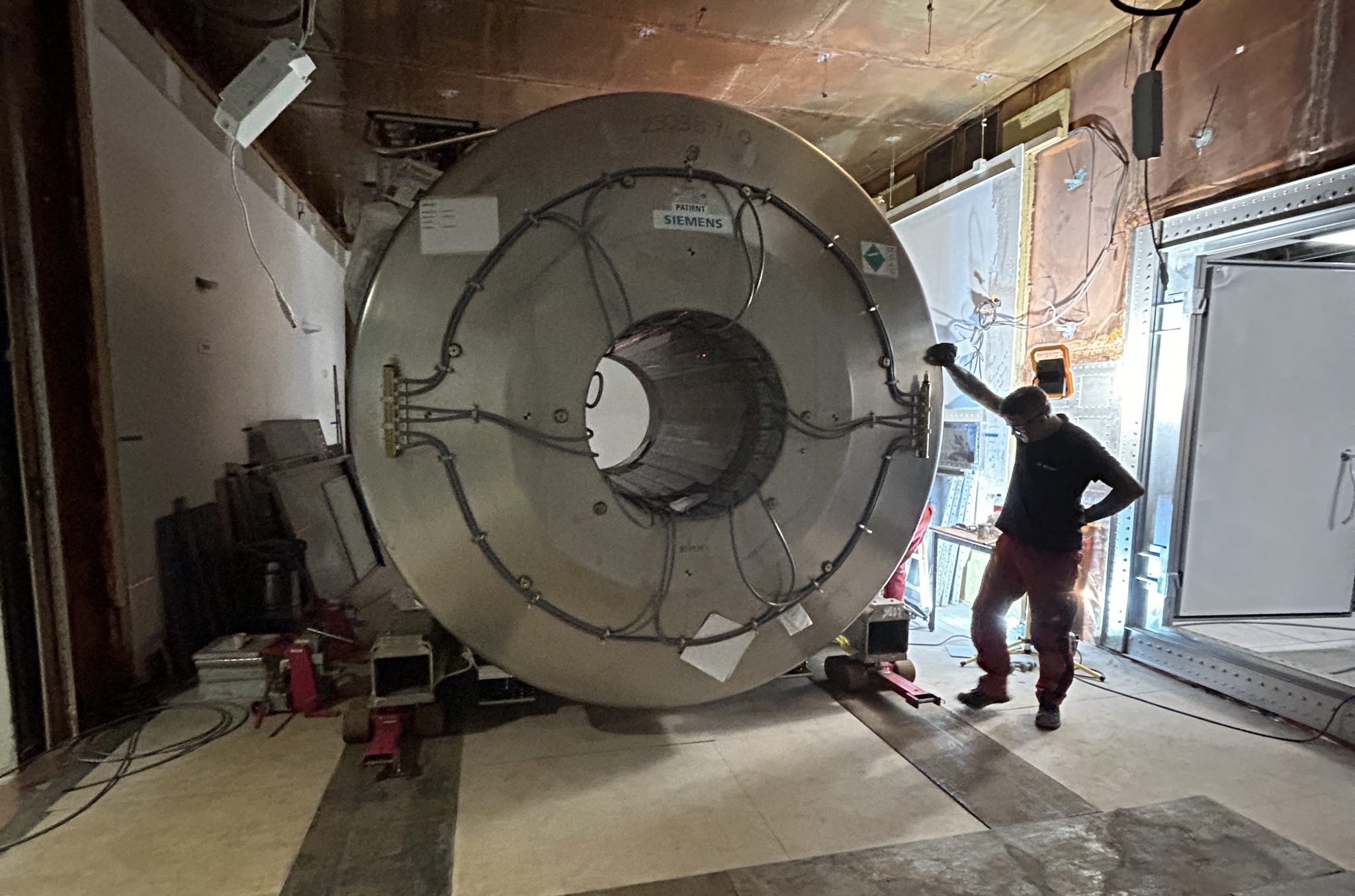
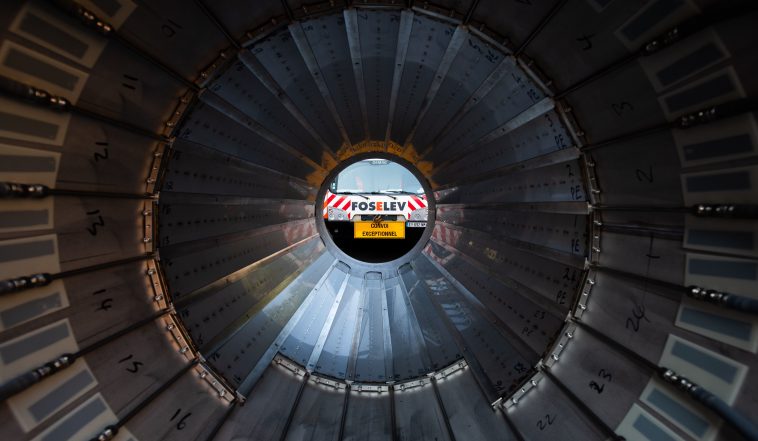
The MRI magnet
As the magnet is increasingly used in the field, as planned for the summer, an adjustment and fine-tuning phase will be necessary. This will require the expertise of staff at the CENIR facility, and an engineer from Siemens Healthineers, who is on full-time secondment to Paris Brain Institute. This adjustment phase will help to optimize how this technology is used for brain and spinal cord exploration.
The first research protocols should begin in early 2025. The expected technological leap comes mainly from increasing the signal-to-noise ratio, which will make it possible to observe submillimeter resolution tissue structures by improving the spatial resolution of the 7T MRI and obtaining images of molecules whose signal is too weak at a lower field, as well as from improving the contrast of images so as to make it possible to visualize structures invisible at weaker magnetic fields.