

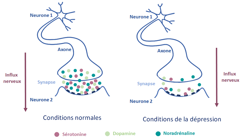


Depression results from a malfunction in the transmission of information from one neuron to another in the brain. Between two neurons there is a gap, called a synapse, where the neuron transmitting the information releases molecules called neurotransmitters, which are captured by the neuron receiving the information. The capture of neurotransmitters by a neuron leads to the generation of an electrical current, the nerve impulse, which travels via the axon, the extension of the neuron, to the next synapse and triggers the secretion of neurotransmitters.
A disturbance in the production and uptake of 3 major neurotransmitters is at the root of the development of a major depressive episode.
- Serotonin, whose function is to balance sleep, appetite and mood
- Dopamine, responsible for regulating mood and motivation
- Noradrenaline, which manages attention and sleep
These neurotransmitters are present in many regions of the brain. Using functional brain imaging (functional MRI), it is possible to identify the brain areas most involved in depression.
Depression is a disorder affecting the dynamics of the brain networks involved in emotional regulation, cognitive control and self-reference.
At the Paris Brain Institute
The ANR “SENSO” project coordinated by Prof. Philippe FOSSATI has made it possible to define brain imaging markers to help diagnose depression. Using functional MRI, it has been possible to identify dysfunction in a network linking different brain regions. Under normal conditions, the role of this neural network is to determine which external stimuli are of interest.
Among other things, this project highlighted the role of the medial prefrontal cortex, which plays a role in sadness and self-deprecation, the left dorsolateral prefrontal cortex, which is involved in working memory, and the precuneus, which is associated with ruminations. He also highlighted the role of the ventral cingulate cortex in sensitivity to signals of social exclusion, a key factor in triggering a major depressive episode.
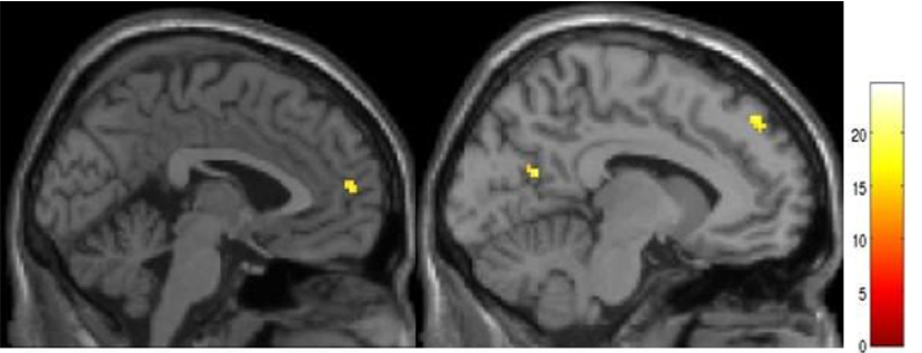
Functional MRI image, activated regions identified in yellow.