

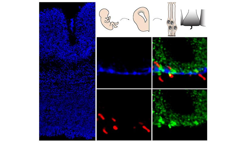
Section of human brain (cortex). On the left, the nuclei are marked in blue; on the right, the cilium at the apical foot (in green) of the progenitor cells is For more information
Section of human brain (cortex). On the left, the nuclei are marked in blue; on the right, the cilium at the apical foot (in green) of the progenitor cells is marked in red. The apical surface is marked in blue.
© Monia Barnat/Grenoble Institut des Neurosciences/Inserm, Université Grenoble Alpes